
Human Eyes and Sight (मानव आँख) – Biology Topic.
Biology topic – “Human Eyes and Sight (मानव आँख)”, is important for all competitive exams like SSC CGL, SSC CHSL, RRB NTPC, UPSC and other state civil services exams. In these exams, almost 4-5 questions are coming from Biology. Let’s start the topic- Human Eyes and Sight (मानव आँख):
Human Eyes and its Sight (मानव आँख)
Eyes: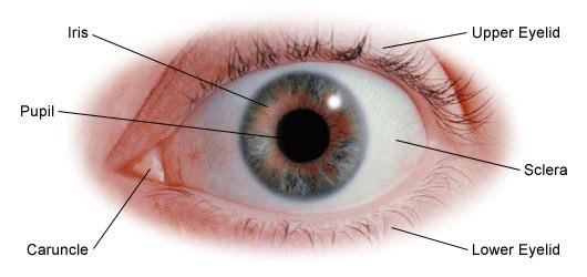
Eyes are organs of the visual system. Our eyes acts like a camera which provide us vision, the ability to receive and process visual details with the help of our brain. They take pictures of the world around us and send the pictures to our brain. Our brain works out what your eyes are seeing. We see through our eyes, which are organs that take in light and images and turn them into electrical impulses that our brain can understand.
Human Eyes Anatomy
Cornea :
This is the layer that covers the front of our eye. It is clear like glass and it has no blood vessels in it. It focuses the light that is coming in through it, and with the lens it makes sure that the image that reaches the back of the eye is in focus.
Sclera :
This is the tough white skin which covers the outside of the eyeball (except for the see-through cornea). We call it the ‘white’ of the eye.
Iris :
The iris controls the amount of light that enters the eye. The iris is the colored part of our eye.
Pupil:
This is the hole in the middle of the colored iris. It lets light into our eye. It gets very small in bright light, and bigger in dull light. The pupil can change sizes with the help of the colored part around it, a muscle called the iris. By opening and closing the pupil, the iris can control the amount of light that enters the eye. If the light is too bright, the pupil will shrink to let in less light and protect the eye. If it’s dark, the iris will open the pupil up so more light can get into the eye.
Retina:
This acts as the sensors in our eyes. The retina turns the light rays into signals that our brain can understand. The retina has two light sensitive cells called ‘rods’ and ‘cones’ (because that is what they look like.) The rods are extra sensitive to light and help us to see when it’s dark. Rods can ‘see’ black and white. The cones help us to see color. There are three types of cones each helping us to see a different color of light: red, green, and blue. They turn the picture into an electrical message for the brain. Sometimes people don’t see all the colors due to ‘Color blindness’.
Blind spot :
This is a bit of our retina which is not sensitive to light because there are no rods or cones there. It is the spot where the optic nerve is joined on to the retina.
Optic nerve :
The electrical messages from the retina travel along the optic nerve to our brain.
Tear glands (Lacrimal Gland) :
These are small glands inside our upper eye-lid known as Lacrimal Gland. Their job is to make tears to keep the surface of our eyeball clean and moist, and help protect your eye from damage.
When we blink, our eyelids spread the tears over the surface of the eye. Mostly the tears flow down a tiny tube at the edge of our lower eyelid, next to our nose. (If you look very carefully you can see a tiny dot that is the beginning of that tube). This tube carries the tears to the back of our nose and that is why our nose ‘runs’ when we cry!
Conjunctiva : (Thinnest skin of the body)
This is the lining on the inside of our eyelid and the outside of the sclera at the front of our eye (except for the special skin of the cornea). There are some tiny blood vessels on the conjunctiva over our eye. If our eyes get sore, these blood vessels get bigger and our eye looks red.
Ciliary Muscles :
These are a circle of tiny muscles around the lens. They change the shape of the lens by squeezing and relaxing. They squeeze (making the lens fat) to look at nearby objects, and relax (making the lens thinner) for far away objects.
The lens :
The lens helps the cornea to focus light onto the retina. It changes shape (getting thinner or thicker) to make sure that the ‘picture’ on the retina is as clear as possible.
There are two lots of fluid inside the eye.
- Aqueous humour:
Aqueous means water, and humour means fluid. This watery fluid fills the front of the eyeball between the cornea and the lens. - Vitreous humour:
This is a thicker jelly-like liquid which fills the larger part of the eyeball behind the lens and keeps it in shape. (Vitreous means glassy, because the vitreous humour is very clear, so that light can pass through it).
Human Eyes and Sight (मानव आँख)
How does our eye work?
Our eye works in a similar way to a camera. When we look at an object, light reflected from the object enters the eyes through the pupil and is focused through the optical components within the eye.
The front of the eye is made of the cornea, iris, pupil and lens, and focuses the image onto the retina. The retina is the light sensitive membrane that covers the back of the eye. This membrane consists of millions of nerve cells which gather together behind the eye to form a large nerve called the optic nerve.
At the front of the eye is the cornea and anterior chamber underneath it. Below that is the iris, the colored ring which surrounds the pupil, which is the opening in the center. The lens is underneath the iris and held in place by the suspensory ligament at the top and the posterior chamber and ciliary body and muscle below the eye. The retina forms the circular shape at the back of the eye surrounded by a layer called the choroid and then another layer called the sclera. Within the retina is the macula the size of a pinpoint, vitreous body and blood vessels. The optic nerve protrudes from the back of the eyeball and consist of blood vessels.
When the light enters the eye, it is focused to a pin-point on the macula, a small area in the center of the retina at the back of the eye. The macula is responsible for central detailed vision, allowing you to see fine detail and color, read and recognize faces.
When light stimulates the nerve cells in the retina, messages are sent along the optic nerve to the brain. The optic nerves from the two eyes join inside the brain. The brain uses information from each optic nerve to combine the vision from the two eyes allowing you to see one image.
Human Eye Diseases and their Solution:
Myopia (निकट दृष्टि दोष): 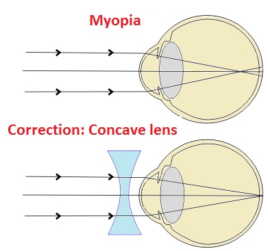
Myopia, otherwise known as shortsightedness or nearsightedness. Nearsightedness (myopia) is a common vision condition in which we can see objects near to us clearly, but objects farther away are blurry. A Person suffering from Myopia has difficulty in vision of farther (Distanced) objects.
- It occurs when image formation of object takes place in front of our retina instead of on the retina.
- It happens due to elongation (increase in size) of Eyeball.
Solution:
It is corrected by the use of Concave (-ve) Lenses (अवतल लेन्स).
Human Eyes and Sight (मानव आँख)
Hypermetropia (दूर-दृष्टी दोष/ दीर्घदृष्टि दोष): 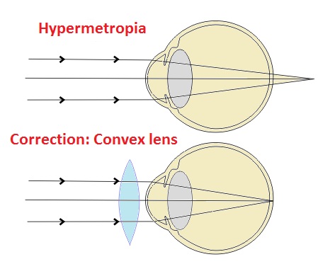
This is also known as Farsightedness. A Person suffering from Hypermetropia has difficulty in vision of nearer object.
- It is a condition of the eye in which light is focused behind the retina, instead of on the retina.
- This results in close objects appearing blurry, while far objects may appear normal.
Cause
- Low converging power of eye lens because of weak action of ciliary muscles
- Abnormal shape of the cornea.
Solution: It is corrected by the use of “Convex Lenses” (+ve).
Presbyopia (जरादूरदृष्टि): 
Presbyopia is the normal loss of near focusing ability that occurs with age. It is an old age eye problem occurs due to weakness of eye muscles.
- Presbyopia is an age-related condition that causes blurred near vision and sometime farther vision as well.
- It typically starts at around age 40.
Solution: It can be corrected by use of both lenses – concave and convex in a single spectical called Bi-focal Lenses. Other Lenses are- trifocals and progressive eyeglasses, used to remove this disorder of eyes.
Color Blindness (Color vision deficiency):
Sometimes a person may not have some types of cone cells. This means that some people cannot see some colors.
- Some people may be unable to see the colors green and red – this is the most common type of color blindness. They might see red as orange and green as white.
- A few people have cones missing which cause them to be unable to see blue and yellow.
How do we get color blindness?
- This is a Genetic Problem. A person gets Color vision deficiency from his parents, and they got it from their parents.
- The gene (जीन) for color vision is on the X chromosome. Females have two X chromosomes and will only be color blind if both those chromosomes are affected. Males have only one X chromosome, so they only need that chromosome to be affected – that’s why color blindness is more common in males.
- Most of the people with color blindness are male – about 1 boy in 10 will be color blind, while only about 1 girl in 200 will be color blind.
Solution: As this is genetics problem, there is no treatment available till now.
Human Eyes and Sight (मानव आँख)
Cataracts / Glaucoma (मोतियाबिंद) :
- Cataract is a change in the lens of the eye; the result is cloudiness as light is prevented from entering the eye properly. It is a clouding of the lens inside the eye which leads to a decrease in vision
- Glaucoma is a condition where a build-up of pressure in the eye causes damage to the optic nerve which is the vital link of the eye to the brain which processes visual information.
Solution : Both of these are corrected by Surgery Only.
Astigmatism (अबिन्दुकता):
- It is an imperfection in the curvature of our eye’s cornea or lens. With astigmatism our vision is blurry at all distances.
- A person suffering from Astigmatism has difficulty in vision of ” Horizontal as well as Vertical” simultaneously.
- It is also called Dimensional Problem of human eye.
Solution:- It can be corrected with the help of required Cylindrical Glasses.
Conjunctivitis:
- Conjunctivitis, is an infection cause irritation or inflammation of the conjunctiva, which covers the white part of the eyeball.
- It can be caused by allergies or a bacterial or viral infection.
- Conjunctivitis can be extremely contagious and is spread by contact with eye secretions from someone who is infected.
Symptoms: Redness, itching and tearing of the eyes. It can also lead to discharge or crusting around the eyes.
Solution: It often resolves on its own, but treatment can speed the recovery process. Allergic conjunctivitis can be treated with antihistamines. Bacterial conjunctivitis can be treated with antibiotic eye drops.
Accommodation (दृष्टी समायोजन) of Vision:
Under this process our near point vision and far point vision should act properly simultaneously. Accommodation is the process by which the vertebrate eye changes optical power to maintain a clear image or focus on an object as its distance varies. In this, distances vary for individuals from the far point—the maximum distance from the eye for which a clear image of an object can be seen, to the near point—the minimum distance for a clear image.
Dioptre (Eye vision Measurement Unit):
- A dioptre (+- D) is a unit of measurement of the optical power of a lens or curved mirror, which is equal to the reciprocal of the focal length measured in metres.
- 1 dioptre = 1 m−1.
- It is a unit of reciprocal length. For example, a 3-dioptre lens brings parallel rays of light to focus at 1⁄3 metre.
- A flat window has an optical power of zero dioptres, and does not cause light to converge or diverge.
Human Eyes and Sight (मानव आँख)
Some Important facts about Human Eye for Competitive Exams:
- Study of Eye is called: Ophthalmology.
- Eye Specialist (Doctor) is known as: Ophthalmologist.
- Different people have different color of eyes due to color of – IRIS (आइरिस).
- Cattle’s eye glowing at night on falling of light due to presence of special membrane: Septum Luciferin. It contain Luciferase enzymes where photo-chemical reaction takes place. This same process of lightning takes place in – Firefly (जुगनू).
- Human Eyes acts as – Convex Lenses (उत्तल लेन्स).
- Foam (Bubbles) of the shop act as – Convex lens.
- In Eye Donation means – Donation of “Cornea”.
- Thinnest skin of human body is – Conjunctiva (eye’s skin).
- Light enters in the eye through the – Pupils (पुतली).
- The size of Pupil is controlled by – Ciliary Muscles.
- Image formed on Retina: Real, Inverted and Diminished in size. (वास्तविक, उल्टी और आकर में छोटा).
- The human eyeball reaches it full size by the age of – 12 years old.
- Tears secreted (स्रावित) from – Lacrimal Gland (लेक्रामल ग्रन्थि).
- Tears are slightly Antiseptics in nature due to presence of – Lysozyme Enzyme.
- Retina is made of two type’s cells – Rod Cells and Cone Cells.
- Crocodile always looks weeping due to absence of Nasal Pharyngeal duct.
- Rod Cell- It control the intensity of light and its all function is regulated by Vitamin – A (Retinol).
- Deficiency of Vitamin – A is cause of – Night Blindness / Xerophthalmia (रतौंधी).
- Cone Cells: control the vision of Colors.
- There are 40-types of Cone-Cells. Hence we can distinguish between (पहचान कर सकतें है) 40 Shades of a Color.
- Human eye can distinguish between 7 X 40 = 280 colors at a time.
- Human eye is most sensitive for – Yellow color. Hence the color of school buses, taxis, PCO and public Advertisement in Yellow Color.
- The power of lenses measured by Dioptre (+/- D).
- Vision distance of 6/6 (correct) eye 1) for near point = 25 C.M. and for farther point = ∞
- The outer layer of the eye (fibrous tunic) contains the cornea and the sclera.
- The middle layer of the eye (vascular tunic) contains the choroid, ciliary body, iris and the pigmented epithelium.
- The innermost layer of the eye contains the retina and blood vessels.
- The human eyes blink between 15 and 20 times/minute. That means you blink up to 1,200 times per day.
- Someone who is shortsighted has an eyeball that is larger than normal and is unable to see things clearly far away.
- Someone who is farsighted has an eyeball that is shorter than average and is unable to see things clearly close-up.
- Approximate 80% of world blindness occurs due to – Cataract disease.
- We have a blind spot where the optical nerve connects to the retina.
- Protanopia is the scientific term for the condition commonly known as red-green color blindness.
- Approx. 8 % of men are color blind, but less than 1 % of women.
- The most common eye color worldwide is – Brown.
For More Click:
If you like and think that topic “Human Eyes and Sight (मानव आँख)” is helpful for you, Please comment us. Your comments/suggestions would be greatly appreciated. Thank you to be here. Regards – Team SukRaj Classes.